What is Respiratory Syncytial Virus Cause Effect Infection?
How does Respiratory Syncytial Virus Infection cause?
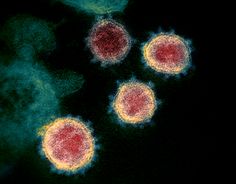
Caused by respiratory syncytial virus or fusion virus (RSV).
It belongs to paramyxovirus RNA type, with a diameter of 100-140nm. The
nucleocapsid is composed of 32 symmetrical icosahedral capsids and has a
capsule. Not destroyed by ether and chloroform. Human cells, diploid cells, and
primary monkey kidney cells can be used to culture viruses and produce special
fusion cells. With fluorescent antibody technology, the virus can be detected
in the cytoplasm of infected cells. The disease is transmitted through the
droplet respiratory tract and has the characteristics of wide spread, high
infection rate, and long duration. It is spread and spread in various countries
around the world, and a major epidemic occurs almost every year or every other
year.
Pathogenesis of Respiratory syncytial virus infection
Respiratory syncytial virus infection passes through air
droplets or directly into the respiratory tract of susceptible persons. After
RSV invades the body, it first multiplies in the nasopharyngeal mucosa and
causes upper respiratory tract infection.
Infants with low immune function, the
elderly, RSV can extend from the nasopharynx to bronchial and alveolar levels
at all levels, and then develop severe bronchitis, bronchiolitis and pneumonia.
Respiratory virus invades human ciliary epithelial cells on
the surface of the respiratory tract, replicates and spreads within it and
directly causes damage to infected cells, causing local lesions or producing
symptoms of systemic toxins.
Some virus-infected tissue damage may be mediated
by the body's immune response. For example, respiratory syncytial virus has the
least direct damage to respiratory cilium epithelial cells, but can cause
serious respiratory diseases in infants and young children; the most vulnerable
age is mother-to-child transmission.
The stage of the highest antibody level
Aafter the
vaccination, the condition of the naturally infected person is worsened, all
suggesting that the onset may be related to the immune response. The
pathological changes of respiratory virus infection include nasal, pharyngeal,
and laryngeal mucosal congestion, edema, exudation, and monocyte infiltration.
Some cells can degenerate, necrotize, and fall off. Inclusion bodies can be
seen in the cytoplasm or nucleus of epithelial cells.
The extent of the disease is related to the type, type and
site of infection. After a few days, the epithelial cells can regenerate and
return to normal. If the lesion involves the bronchioles, epithelial cell
necrosis and exfoliation can occur. The bronchiolar wall has extensive
mononuclear cell infiltration. Fibrin, cell debris and thick mucus can block
the lumen and cause atelectasis and emphysema.
Viral Pneumonia Manifestation
Viral pneumonia initially manifests as a progressive
reduction of cilia, formation of vacuoles in epithelial cells, followed by
degeneration of epithelial cells, substantial necrosis and collapse of alveoli,
necrosis and thickening of alveolar walls, interstitial edema and monocytes,
lymphocytes infiltration.
When bacterial infection is complicated, mucosal
hyperemia, neutrophil infiltration, and mucopurulent secretion can be seen. In
severe cases, pulmonary abscess, sepsis and purulent changes in multiple
organs can occur.
Share on Social Media to Help Someone who may be in Need of this Info >>


